Semi-Quantitative Cell-Based Indirect Fluorescent Antibody / Semi-Quantitative Indirect Fluorescent Antibody (IFA) / Qualitative Immunoblot / Semi-Quantitative Enzyme-Linked Immunosorbent Assay (ELISA) / Quantitative Radioimmunoassay (RIA)
Semi-Quantitative Cell-Based Indirect Fluorescent Antibody / Semi-Quantitative Indirect Fluorescent Antibody (IFA) / Qualitative Immunoblot / Semi-Quantitative Enzyme-Linked Immunosorbent Assay (ELISA)
Autoimmune movement disorders encompass a large, diverse group of neurologic disorders that can occur in isolation or in conjunction with other autoimmune encephalitides. Detection of antineural antibodies may help to establish a diagnosis, support treatment decisions, aid with prognostication, and guide the search for an associated malignancy.
Disease Overview
Autoimmune movement disorders may resemble genetic, metabolic, or neurodegenerative movement disorders. Importantly, autoimmune movement disorders may be treatable if identified in a timely manner; they may be the presenting symptom of an undiagnosed malignancy, and recognition of tumor-antibody associations may allow for cancer treatment at an early stage. Signs and symptoms associated with these disorders are diverse and may include tremor, ataxia, chorea, neuromyotonia, myokymia, dystonia, myoclonus, abnormal eye movements, and/or parkinsonism. Patients may also have symptoms such as headache, psychosis, hallucinations, and/or agitation. Subacute onset of symptoms, inflammatory cerebrospinal fluid (CSF) studies, and magnetic resonance imaging (MRI) findings consistent with inflammation or cerebellar degeneration may support this diagnosis.
For more information about laboratory testing for autoimmune neurologic diseases, refer to the ARUP Consult Autoimmune Neurologic Diseases - Antineural Antibody Testing topic.
Test Description
These serum and CSF antineural antibody panel tests may be used for the evaluation of patients with subacute onset of movement disorders. Testing for the presence of antineural antibodies in both serum and CSF may improve diagnostic yield.
These phenotype-targeted panels test for the presence of antibodies associated with movement disorders. Clinical phenotypes for specific antineural antibody-associated syndromes often overlap, and phenotype-specific panels allow for rapid identification of associated antibodies, which may have implications for treatment, prognosis, and cancer screening. Other panels may be more appropriate, depending on the patient’s clinical phenotype:
| ARUP Panel | Test Code | |
|---|---|---|
| Serum | CSF | |
| Autoimmune Encephalopathy/Dementia Panel | 3006201 | 3006202 |
| Autoimmune Epilepsy Panel | 3006204 | 3006205 |
| Autoimmune Pediatric CNS Disorders Panel | 3006210 | 3006211 |
Regardless of the panel chosen, order only one panel for serum and/or one panel for CSF; many antineural antibodies are redundant between these panels, and choosing based on the predominant phenotype will provide the most meaningful results. To compare these panels and the antibodies included, refer to the ARUP Antineural Antibody Testing for Autoimmune Neurologic Disease page.
Testing for individual autoantibodies is also available separately.
Antibodies Tested and Methodology
| Autoantibody Markers | Methodology | Individual Autoantibody Test Code | |
|---|---|---|---|
| Serum | CSF | ||
| AMPAR Ab, IgG | CBA-IFA, reflex titer | 3001260 | 3001257 |
| Amphiphysin Ab, IgG | IB | 2008893 | 3004510 |
| ANNA-1 (Hu) | IFA, reflex IB, reflex titer | 2007961 | 2010841 |
| ANNA-2 (Ri) | IFA, reflex IB, reflex titer | 2007961 | 2010841 |
| CASPR2 Ab, IgG | CBA-IFA, reflex titer | 2009452 | 3001986 |
| CV2 (CRMP-5) Ab, IgG | CBA-IFA, reflex titer | 3016999 | 3017001 |
| DPPX Ab, IgG | CBA-IFA, reflex titer | 3004359 | 3004512 |
| GABA-AR Ab, IgG | CBA-IFA, reflex titer | 3006008 | 3006003 |
| GABA-BR Ab, IgG | CBA-IFA, reflex titer | 3001270 | 3001267 |
| GAD65 Ab | ELISA | 2001771 | 3002788 |
| IgLON5 Ab, IgG | CBA-IFA, reflex titer | 3006018 | 3006013 |
| ITPR1 Ab, IgG | CBA-IFA, reflex titer | 3006031 | 3006023 |
| Kelch-Like Protein 11 | CBA-IFA, reflex titer | 3018507 | 3018508 |
| LGI1 Ab, IgG | CBA-IFA, reflex titer | 2009456 | 3001992 |
| Ma2/Ta Ab, IgG | IB | 3017441 | 3017440 |
| mGluR1 Ab, IgG | CBA-IFA, reflex titer | 3006044 | 3006039 |
| NMDAR Ab, IgG | CBA-IFA | 2004221 | 2005164 |
| PCCA-1 (Yo) | IFA, reflex IB, reflex titer | 2007961 | 2010841 |
| PCCA-Tr/DNER | IFA, reflex IB, reflex titer | 2007961 | 2010841 |
| P/Q-type VGCC Ab, IgG | RIA | 0092628 | — |
| SOX1 (AGNA) Ab, IgG | IB | 3002885 | 3002886 |
| Ab, antibody; AGNA, antiglial nuclear antibody; AMPAR, alpha-amino-3-hydroxy-5-methyl-4-isoxazolepropionic acid receptor; ANNA-1, antineuronal nuclear antibody type 1; ANNA-2, antineuronal nuclear antibody type 2; CASPR2, contactin-associated protein 2; CBA, cell-binding assay/cell-based assay; CRMP-5, collapsin response-mediator protein 5; DNER, Delta/notch-like epidermal growth factor-related receptor; DPPX, dipeptidyl-aminopeptidase-like protein 6; ELISA, enzyme-linked immunosorbent assay; GABA-AR, gamma-aminobutyric acid receptor, type A; GABA-BR, gamma-aminobutyric acid receptor, type B; GAD65, glutamic acid decarboxylase 65-kd isoform; IB, immunoblot; IFA, indirect immunofluorescence assay; IgLON5, IgLON family member 5; ITPR1, inositol 1,4,5-trisphosphate receptor type 1; LGl1, leucine-rich, glioma-inactivated protein 1; mGluR1, metabotropic glutamate receptor 1; NMDAR, N-methyl-D-aspartate receptor; PCCA-1, Purkinje cell cytoplasmic antibody type 1; PCCA-Tr, Purkinje cell cytoplasmic antibody type Tr; RIA, radioimmunoassay; SOX1, SRY-box transcription factor 1; VGCC, voltage-gated calcium channel | |||
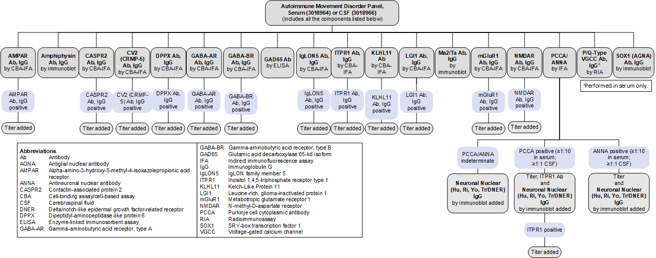
Reflex Patterns
Autoimmune Movement Disorder Panel, Serum (3018964) and CSF (3018966): Reflex Patterns

Limitations
These panels do not include every antibody that has been associated with autoimmune movement disorders:
- ANNA-3 and PCCA-2 are not included because they are extremely rare (present in approximately 0.0001% of specimens submitted for evaluation using a paraneoplastic antibody panel), and commercial assays to confirm the specificity of these antibodies are not currently available.
- Adaptor protein 3 subunit B2 (AP3B2), glial fibrillary acidic protein (GFAP), GTPase regulator associated with focal adhesion kinase 1 (GRAF1), neuronal intermediate filament (NIF) and its associated reflexes (NIF heavy and light chain, alpha internexin), neurochondrin, septin 5, and septin 7 antibodies are not included because they have been only recently identified and their prevalence is currently not well established.
- GFAP has been reported in 0.17% of samples screened, often co-occurring with other antineural antibodies.
- GRAF1 has only been described in rare case reports; its prevalence remains unknown.
- NIF has been reported in 0.014% of samples screened; NIF heavy and light chain and alpha internexin were reflexed in samples that were positive for NIF to further identify the associated antibody.
- Neurochondrin has been reported in 0.002% of samples tested.
- Septin 5 has been reported in <0.001% of samples screened.
- Septin 7 has been reported in 0.002% of samples screened.
- As testing for newly described antibodies becomes available and their clinical relevance is established, these panels will evolve to reflect these discoveries.
Test Interpretation
Results
Results must be interpreted in the clinical context of the individual patient; test results (positive or negative) should not supersede clinical judgment.
| Result | Interpretation |
|---|---|
| Positive for ≥1 autoantibodies | Autoantibody(ies) detected Supports a clinical diagnosis of an autoimmune movement disorder Consider a focused search for malignancy based on antibody-tumor associations |
| Negative | No autoantibodies detected A diagnosis of an autoimmune movement disorder is not excluded |
References
-
33324912
Gövert F, Leypoldt F, Junker R, et al. Antibody-related movement disorders – a comprehensive review of phenotype-autoantibody correlations and a guide to testing. Neurological Res Pract. 2020;2:6.
-
34489848
Garza M, Piquet AL. Update in autoimmune movement disorders: newly described antigen targets in autoimmune and paraneoplastic cerebellar ataxia. Front Neurol. 2021;12:683048.
-
27112681
Flanagan EP, Drubach DA, Boeve BF. Autoimmune dementia and encephalopathy. Handb Clin Neurology. 2016;133:247-267.
-
24833664
Horta ES, Lennon VA, Lachance DH, et al. Neural autoantibody clusters aid diagnosis of cancer. Clin Cancer Res. 2014;20(14):3862-3869.
-
29293273
Dubey D, Pittock SJ, Kelly CR, et al. Autoimmune encephalitis epidemiology and a comparison to infectious encephalitis. Ann Neurol. 2018;83(1):166-177.
-
30158896
Bartels F, Prüss H, Finke C. Anti-ARHGAP26 autoantibodies are associated with isolated cognitive impairment. Front Neurol. 2018;9:656.
-
30282771
Basal E, Zalewski N, Kryzer TJ, et al. Paraneoplastic neuronal intermediate filament autoimmunity. Neurology. 2018;91(18):e1677-e1689.
-
31511329
Shelly S, Kryzer TJ, Komorowski L, et al. Neurochondrin neurological autoimmunity. Neurol Neuroimmunol Neuroinflamm. 2019;6(6):e612.
-
36053822
Hinson SR, Honorat JA, Grund EM, et al. Septin‐5 and ‐7‐IgGs: neurologic, serologic, and pathophysiologic characteristics. Ann Neurol. 2022;92(6):1090-1101.


 Feedback